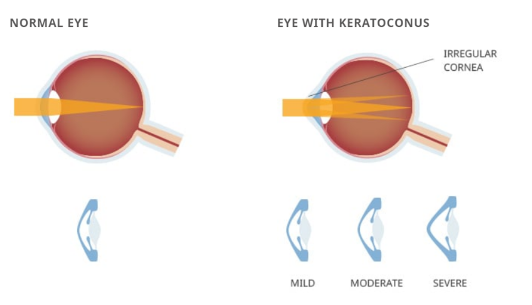
What is Keratoconus?
Keratoconus is a progressive eye disease in which the normally round, dome shaped cornea (the clear outer front portion of the eye) thins and begins to bulge into a cone-like shape. This cone shape is irregular, bending light as it enters the eye, causing distorted vision. Keratoconus can occur in one or both eyes and often begins during a person’s teens or early 20s.
Blurred vision caused by keratoconus (especially in its late stages) is different from blurred vision that is caused by other common refractive disorders such as myopia or hyperopia. In the later stages of keratoconus, achieving satisfactory vision, even with glasses or, contacts, can be difficult and refractive surgery is not recommended. Common refractive errors by comparison are easily correctable.

Signs and Symptoms
To patients, early indications of keratoconus are usually a blurring and distortion of vision. In the early stages this may be barely noticeable, and only specific tests may uncover this condition. The need to frequently change glasses or contacts due to irregular shifts in astigmatism, changes in the curvature of the eye, or thickness of the cornea may be an indication that more specific tests are required. Those with an evolving or progressive case will likely experience the effects gradually over a few years. Occasionally, the thinning can progress more rapidly.
In the advanced stages of keratoconus, the patient may experience a sudden clouding of vision in one eye that clears over a period of weeks or months. This is called “acute hydrops” and is due to the sudden infusion of fluid into the stretched cornea. In advanced cases, superficial scars form at the apex of the corneal bulge resulting in more vision impairment.
