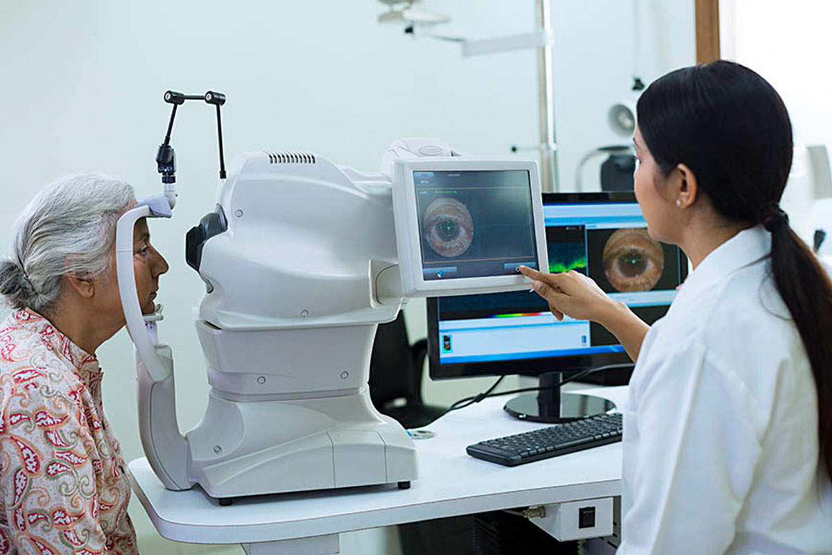
MIGS stands for minimally invasive glaucoma surgery, and refers to a range of implants, devices and techniques which all aim to reduce the pressure in the eye (intraocular pressure, or IOP). Glaucoma is caused by the build-up of a fluid in the eye called aqueous humour. This causes IOP to increase, which damages the optic nerve and leads to vision loss. MIGS improves the drainage of the fluid out of the eye.
“Minimally invasive” means that they use tiny incisions and/or microscopic equipment, which have been designed to reduce risk compared to procedures such as a trabeculectomy or aqueous shunt.
Types of Minimally Invasive Glaucoma Surgery
Below is a list of different minimally invasive glaucoma surgery options available at Hashmanis Group of Hospitals.
- Ab-interno Canaloplasty with iTrack
The technique is not as tedious and complicated as the traditional canaloplasty and can be done by any skilled cataract surgeon. By far, it is consider to be the most promising, of the microinvasive glaucoma surgeries.
Canaloplasty uses an iTrack microcatheter to open the eye’s natural drainage system (“Schlemm’s canal”). Whereas, with traditional Canaloplasty, the Micro-catheter is inserted from an external approach: cutting through the conjuctiva and sclera, while in Ab-Interno Canaloplasty, the Micro-catheter is inserted from an internal approach: through either a clear corneal or a limbal micro-incision, then through a small opening in the Trabecular Meshwork, and into the Schlemm’s canal. With Ab-Interno, the eye tissues are preserved by not cutting the flap.
The micro-catheter is then routed 360° through the Schlemm’s canal. Once the end of the catheter has circumnavigated to the point of entry, the catheter tip is slowly pulled back while sterile, viscoelastic gel is injected through the catheter and delivered gradually along the canal to dilate it 2-3 times its normal size. Unlike with traditional canaloplasty, this enlarges and breaks down obstructions and adhesion in the canal and restores the natural outflow pathways, without leaving a prolene suture in the Schlemm’s canal. Once the outflow pathway is improved, the eye’s fluid can exit through a more natural process allowing the pressure in the eye to drop to a more normal level.
Ab-Interno Canaloplasty Benefits
To date, Ab-Interno Canaloplasty is the only Microinvasive Glaucoma Surgery procedure that successfully addresses all aspects of potential outflow resistance. You get Trabecular Meshwork treated (an opening is created with Ab-Interno), Schlemm’s canal is dilated and Collector channel systems are also re-opened. Hence, you get better aqueous outflow.
- Endocytophotocoagulation (ECP)
ECP is a laser probe which targets the part of the eye which produces the fluid in the eye, called the ciliary body. The laser reduces the activity of the ciliary body, meaning less fluid is produced which reduces IOP. The effect is temporary as the ciliary body eventually recovers, but the treatment can be repeated.
- Hydrus
The Hydrus is an eyelash-sized device which is put into the main drainage channel of the eye (called Schlemm’s Canal). It acts as a scaffold to hold the drainage channel open and allow the fluid in the eye to drain, reducing IOP. It is made of nickel and aluminum.
- iStent
iStents are used as a treatment for glaucoma and, in children, NLD (Nasolacrimal duct) obstruction. Stents are very small tubes usually made of plastic, fabric, or metal, which are surgically inserted to relieve obstructions and keep a path open so blood or other fluids can pass. Stenting to help treat glaucoma is used in combination with cataract surgery to reduce pressure inside the eye in certain adult patients who have mild or moderate open-angle glaucoma. Clear fluid in a healthy eye flows smoothly through the front chamber of the eye and empties out through a mesh of tissue and exits via a canal at the edge of iris and cornea. If the meshwork becomes blocked or drains slowly, pressure builds up inside the eye to a level that may cause vision loss. A stent creates an opening to allow better drainage. In a healthy child, the nasolacrimal duct lets tears pass through the eye by clearing out into the nasal passages. In NLD, that duct is blocked or did not open at birth. NLD obstruction usually opens spontaneously by age 12 months. If it cannot be treated with other therapies, if anatomic abnormalities are present or the obstruction is tight, a silicone stent may be placed with the child under general anesthesia.
- PreserFlo MicroShunt (previously known as InnFocus MicroShunt)
The PreserFlo MicroShunt is a small tube that is inserted into the eye to create a new drainage pathway for the fluid. It acts as a bypass, with the excess fluid draining to a small blister (called a bleb) under the conjunctiva (the surface of the eye), under the upper eyelid. This improves drainage of the fluid in the eye, reducing IOP. It is made of a type of plastic.
- Trabectome
A trabectome uses an electrical pulse to remove part of the blocked drainage channel. This helps the eye’s natural drainage pathway to start working again, allowing the fluid in the eye to drain and reducing IOP.
- XEN Gel Implant
The XEN Gel Implant (pronounced ‘zen’) is a thin tube that is inserted into the eye to create a new drainage path for the fluid. It acts as a bypass, with the excess fluid draining to a small blister (called a bleb) under the conjunctiva (the surface of the eye), under the upper eyelid. This improves drainage of the fluid in the eye, reducing IOP.
- CyPass Micro-Stent
The CyPass Micro-Stent is a very small tube which is inserted to bypass the eye’s main drainage channel and to improve another natural drainage pathway. This improves the drainage of fluid out of the eye, reducing IOP. It is made of a special plastic.
The CyPass was withdrawn in August 2018, as research indicated people with a CyPass had an increased risk of losing cells from the cornea (the surface of the eye). The device is no longer being used but it’s unlikely that people with a CyPass will have it removed, as this is likely to cause damage to the eye.
- Drainage Implant Surgery
Glaucoma drainage implants are small prosthetic devices that are placed to help lower the intraocular pressure and prevent further optic nerve damage. Glaucoma drainage implant surgery is an alternative to traditional filtration surgery (Trabeculectomy). In some patients, particularly those with certain types of glaucoma such as Aphakic glaucoma, Neovascular glaucoma, and Uveitic glaucoma, Trabeculectomies are known to be less successful at reducing intraocular pressure due to an aggressive healing response. Also, in patients who have had other eye surgeries, a glaucoma drainage device often works better than a trabeculectomy procedure to control the intraocular pressure. It should be noted that the glaucoma implant is not used to improve vision, but rather to lower intraocular pressure and prevent further vision loss from glaucoma. In this respect, this implant is completely different from the type of implant used during cataract surgery.
Glaucoma drainage implants are also successfully used as an initial surgical procedure for glaucoma. Various factors may influence the surgery recommended by your doctor. Sometimes an implant is necessary because there is expected to be extensive scarring in the outer layers of the eye. Compared to the channel made with Trabeculectomy, the tube of a glaucoma implant is less likely to become blocked by this scar tissue.
How do drainage implants work?
Glaucoma drainage implants come in different shapes and sizes. There are two general types of implants:
Valved and Non-valved implants. All these implants have a tube and plate design. Regardless of which type of implant is used, a silicone tube is inserted into the front of the eye, usually between the cornea and iris, but other locations are occasionally used. The tube is like an artificial drain, allowing fluid to pass through it to a plate, which has been placed on the surface of the eye and acts as a reservoir.
The fluid then slowly percolates through this reservoir and is absorbed into the body fluids. The implant plate is usually placed in the area underneath the upper eyelid. Unless the lid is pulled back, neither you nor your family will notice it. With the upper lid retracted, a clear or white patch may be noted. This is a patch that covers the tube and prevents irritation. With all drainage implants, it can take 3 months or longer after surgery for the intraocular pressure to stabilize, as the capsule surrounding the plate of the implant needs time to mature in the eye.
What is my chance of success with after Drainage Implant Surgery?
Studies have shown that the success of glaucoma drainage implants is similar to those of trabeculectomy. It should be noted that glaucoma implants are sometimes used in patients with more complicated problems, and therefore the success rate in these patients may be lower than trabeculectomy in a standard eye. However, in many patients, these implants may be the best remaining available option. In about 5-10% of cases a second tube implant is necessary to adequately control intraocular pressure. When a second tube is necessary it is usually place in the lower part of the eye under the lower eyelid.
Remember that the goal of glaucoma implant surgery is to lower intraocular pressure and preserve vision. It will not restore vision that has already been lost. By lowering eye pressure, it is hoped that the operated eye will be spared further glaucomatous damage and can maintain its vision. As with any eye surgery, there is a risk of loss of vision, though this risk is low. Sometimes your doctor will combine the tube implant surgery with cataract surgery. In these cases there may be some visual improvement from clearing of the cataract and replacing it with a clear intraocular lens implant.
What is involved with a Glaucoma tube procedure?
After discussing the risk, benefits, and alternatives to surgery, your doctor will decide on the appropriate type of tube implant to be placed in your eye. When you and your doctor make a decision to proceed with placement of a glaucoma drainage implant, you will meet with our pre-operative counsellor who will give you detailed instructions on how to prepare yourself for your upcoming surgery and what is involved in getting to the operating room for the procedure.
In most cases, the surgery takes about one hour, though you will be at the surgery center for about 3-4 hours. The surgery is usually done under local anesthesia with intravenous sedation. An injection of local anesthetic numbs the eye completely so there is no discomfort and the eye will not move during surgery. Uncommonly a general anesthetic is used and the patient is put to sleep for the operation. Local anesthesia offers several advantages including less pain post-operatively, no sore throat from the airway tube used in general anesthesia, and quickly returning to normal alertness without the nausea often felt after general anesthesia. With local anesthesia, there is less risk than with a general anesthetic, especially in the elderly or those with health problems.
After surgery, the eye is covered by an eye patch and protected by a plastic shield overnight. On the morning following surgery, the patch/shield is removed and your ophthalmologist examines the eye. Eye drops are then used to prevent infection and reduce inflammation. It is important to take these as directed by your ophthalmologist since they can make a great deal of difference in the success of the procedure.
It may take several months after your surgery for the healing to be complete and for the implant to mature in your eye. During this time it is not unusual for your intraocular pressure, as well as your vision, to fluctuate. You will be ready to change your glasses prescription approximately 2-3 months after surgery.

